They can rot, crack, weaken, become mushy, and eventually peel away and become brittle. Remember, too, that thick fences and drywall can absorb material such as sand, dirt, dust, paint, water, mold, etc. A damaging hole would be created in the drywall or wooden structure, Gram-positive bacteria in this analogy. However, if someone shot a bullet at a thick wooden fence, or shot through a drywalled barrier in a room, the projectile would probably penetrate these surfaces and blast completely through. Thus, in this analogy, our Gram-negative bacteria (the person wearing the bulletproof vest) would likely be unharmed. The bulletproof vest is difficult to penetrate with powerful weapons. If someone used a common gun and shot a slug at the bulletproof vest, it would probably not penetrate or go through it. Remember, a bulletproof vest is very thin, while a heavy wood fence or a drywalled partition is quite thick. How do Gram-negative and Gram-positive bacteria influence bacterial evolution and natural health? Meet Sian Seligman: ACHS Grad Making Waves in the Plant Medicine Field
Gram positive vs gram negative color free#
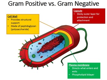
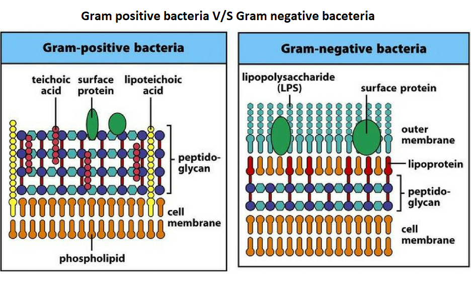
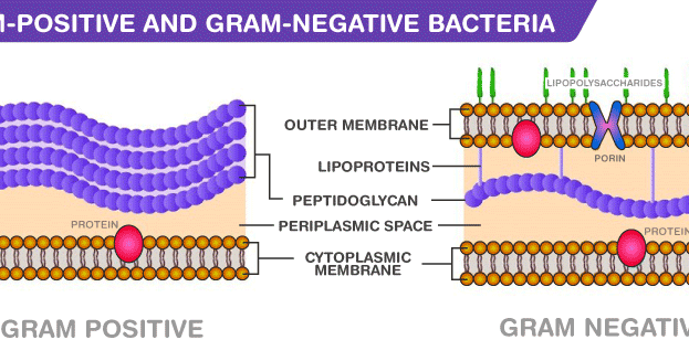
Image copyright free from Wikimedia Commons at Image: Structure of gram-negative cell wall. This thick outer covering, or membrane, is capable of absorbing a lot of foreign material. Gram-positive bacteria have a greater volume of peptidoglycan (a polymer of amino acids and sugars that create the cell wall of all bacteria in their cell membranes), which is what makes the thick outer covering.

Some examples of Gram-positive bacteria include Streptococcus, Staphylococcus, and Clostridium botulinum (botulism toxin). The cell membrane of Gram-positive bacteria can be as much as 20-fold thicker than the protective covering of Gram-negative bacteria. What do natural health professionals need to know about Gram-positive bacteria? The protective covering of these, and other, Gram-negative bacteria make them much more difficult to heal and eradicate. Examples of Gram-negative bacteria include cholera, gonorrhea, and Escherichia coli ( E. Because of this nearly “bulletproof” membrane, they are often resistant to antibiotics and other antibacterial interventions. Gram-negative bacteria’s cell membrane is thin but difficult to penetrate. Image is copyright free from Wikimedia Commons at What do natural health professionals need to know about Gram-negative bacteria? Image: Structure of Gram-positive cell wall. Gram-negative bacteria have a thin membrane, which is nearly “bulletproof.” Gram-positive bacteria have a big, thick membrane. The key to understanding these differences is in the protective membrane, or outer covering, surrounding these bacterial organisms. Janet Carter: Empowering Veterans Through Herbal Medicine
Gram positive vs gram negative color registration#


Decolorizing the cell causes this thick cell wall to dehydrate and shrink, which closes the pores in the cell wall and prevents the stain from exiting the cell. The cell walls of gram positive bacteria have a thick layer of protein-sugar complexes called peptidoglycan and lipid content is low. When the bacteria is stained with primary stain Crystal Violet and fixed by the mordant, some of the bacteria are able to retain the primary stain and some are decolorized by alcohol. This test differentiate the bacteria into Gram Positive and Gram Negative Bacteria, which helps in the classification and differentiations of microorganisms. Gram Staining is the common, important, and most used differential staining techniques in microbiology, which was introduced by Danish Bacteriologist Hans Christian Gram in 1884.


 0 kommentar(er)
